I n addition, mechanical mismatch between soft-tissue and metal leads to stress shielding at the tissue interface, hampering load transfer across the graft and promotes tissue damage and implant loosening. Buildup of particles around the materials due to wear would potentially incite inflammatory response and activate osteoclasts thereby triggering the osteolysis and other associated hypersensitivity reactions. These concerns demand revision surgery for the removal of conventional metallic screws. However, surgical removal of the conventional metallic screws after healing is associated with undesirable complications such as painful morbidity, removal of bone tissue if any, oseointegration and screw breakage.
Emergence of biodegradable screws promises the gradual dissolution and metabolization of implants by the body and thereby overcoming the limitations of metallic screws. Biodegradable screws are potential substitutes that replace metallic screws as it does not require any surgical excision. Synthetic polymers such as polylactide, polyglycolide and poly (lactide-co-glycolide) are widely used in the fabrication of syndesmotic screws.
These biodegradable screws have been found to reduce the stress shielding effect as it gradually transfers the load to the healing tissue while it resorbs. Though these polymeric screws show minimal interference of MR imaging, implantation of these screws has been associated with many clinical complications such as screw fracture, sterile abscess formation, and acute inflammation with the screw displacement due to the lack of host-tissue integration and accumulation of acidic degradation products. Poor integration with surrounding host tissue is mainly due to the lack of any bioactive signals in these synthetic biomaterials.
The pathological complications of biodegradable screws obviate the surgical removal of polymeric screw fragments. Hence, this Indo-U.S. Joint Center on
“Orthopedic Regeneration” is currently working in the development of interconnected porous biodegradable orthopedic screws for syndesmotic injuries. We hypothesized that the open porosity of the screws helps to immigrate the healthy cells from the surround tissue. The tissue surrounding the screws become fragile leading to localized osteopenia due to the employment of physical force during implantation. Thus the open porous structures of these screws would facilitate the mechanical interlocking of the weak surrounding tissue with the screws that would directly avoid the screw loosening. Fabricating screws with open porous structures using traditional fabrication techniques like compression molding and machining are tricky. Hence, 3D printing technology has been employed for large scale manufacturing of biodegradable cortical screws for surgical implantation in syndesmotic injuries. For the first time, porous biodegradable screws have been fabricated with tunable porosity promising high reproducibility for complex porous architecture thus maintaining a balance between function and mass transport. This additive manufacturing based porous biodegradable screw designs would avoid the existing biological complications of polymeric screws by exhibiting excellent oseointegration along with neo-vascularization.
Indo-U.S. Joint Center for Orthopaedic Regeneration
The primary focus of this partnership is to develop personalized orthopedic implants with the ideal chemical and biological cues using 3D printing technology, which would essentially enhance the healing rate. This Joint Center helps the researchers in both countries to move towards the personalization of the implant by sharing newer technologies and process development on either side. Further, the Joint Center allows the partnering investigators to go beyond the primary focus of their individual research to address important areas of biomedical research, education, work force development, and cultural exchange. The center intends to build on long standing collaborations between the partnering investigators, and will leverage complimentary expertise and combined infrastructure to meet the proposed biomaterial challenge.
Our collective work in the area of personalized orthopedic implants has shown the benefits of 3D printing process; design precision, synthesis of semi-synthetic polymer, interconnected porous architecture enabled the biomimicry in maintaining the adequate mechanical properties and vascularization without eliciting inflammatory responses. Indian researchers are involved in the development of biodegradable orthopaedic screws using 3D printing technology.
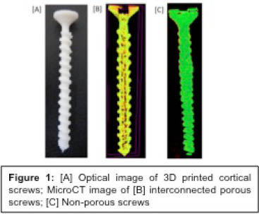
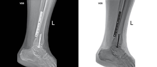
Figure 1 shows the optical image of 3D printed biodegradable screw and the microCT image confirms the interconnected porosity of the screws. Histopathological observations followed by subcutaneous implantation of screws showed no local irritation and observed the presence of minimal inflammatory cells. Interestingly, higher neo-vascularization was seen in tissues implanted with porous screws while there was no evidence of neo-vascularization in non-porous screws. The extent of neo-vascularisation is strongly related to osteogenesis, which is proved in literatures, where neo-vascularisation initiates bone remodeling and repair.
Use of 3D printing technology in the development of orthopaedic screws accomplishes the ease of fabrication, good reproducibility, use of versatile biomaterials and tunable porous architecture. Different head designs were evaluated for enhancing the structural and torsional stability of screws to avoid easy breakage and larger head region above implanted surface that causes microfractures in bones. A better head design was identified as a part of the project to achieve congruous fit while fixations at inclined positions. Furthermore, developing polymer-processing methodologies for semi-synthetic biomaterials may result in new polymeric materials with the ideal chemical and biological properties for 3-D printing.
Scientists from the U.S. are working on the synthesis of newer semi-synthetic polymers and the in vitro characterization of orthopaedic screws. U.S. counterparts recently filed a patent disclosure on the novel bio-erodible cellulose derivative proposed in this study includes 1-oxo-1H-pyrido[2,1-b][1,3]benzoxazole-3-carboxylic acid (AMP) a highly fluorescent molecule as an ionic group to facilitate ionic conductance in the biologic environment. The unusual fluorescent activity of AMP is attributed to additional pair of electrons on the heteroatoms such as oxygen and nitrogen and their circulation around π electrons. This bio-erodible polymer measured a stable ionic conductivity (IC) of 0.12 S/cm up to 6 weeks in vitro at pH 7.4 and 37°C which is significantly higher than many other reports using hydrogels. Additionally, scaffolds derived from this material can be imaged though skin by near infrared imaging (wavelength ~550nm) for the scaffold tracking purposes following its implantation. This AMP-biopolymer will degrade by the cleavage of AMP-ester linkage and cellulose acetate (CA) erodes from the site of implantation via hydrolytic and enzymatic means in in the physiologic environment.
In summary, this Virtual Networked Center is working towards the newer generation of biomaterials and the new knowledge generated via this collaborative program will be extremely useful to develop integrated approaches that harness physical, chemical and biological cues for the development of personalized implants.